Functions and Parts of the Digestive and Urinary Systems
| ✓ Paper Type: Free Assignment | ✓ Study Level: University / Undergraduate |
| ✓ Wordcount: 8763 words | ✓ Published: 06 Jun 2019 |
Table of Contents
i) Parts of the Digestive System
ii) Functions of Digestive System
Introduction
Digestive and Urinary Systems
Part A
The Digestive System consists of a group of organs that work together to change food into energy along with basic nutrients that feed the whole body. It breaks down nutrients into parts small enough for the body to absorb them. Waste products are expelled from the body as faeces. Digestion starts at the mouth and the process ends at the anus. The Digestive System co-ordinates the movement of food together with cells that produce hormones and enzymes which aids the breakdown of food.
Hormones
Hormones are chemical messengers secreted by glands directly into the bloodstream and travel to the target organs (the organ that’s requires them). They influence metabolism, reproduction, growth and development.
Enzymes
Enzymes are proteins which speed up chemical reactions in the body. They breakdown food into small particles so that they can be digested and absorbed by the body.
Organs of the Digestive System
The organs of the digestive system can be divided into those of the (1.) Gastrointestinal Tract and (2.) Accessory Organs.
- The Gastrointestinal Tract is also known as the Alimentary Canal. It is made up of hollow organs as follows;
- Mouth
- Pharynx
- Oesophagus
- Stomach
- Small Intestine
- Large Intestine
- The Accessory Organs also play a vital role in helping your body digest food. The accessory organs are as follows;
- Teeth
- Tongue
- Salivary Glands
- Gallbladder
- Liver
- Pancreas
Functions of the Digestive System
The main functions of the Digestive System include the following processes:
- Ingestion – taking food into the Gastrointestinal Tract
- Propulsion – moves the food along the digestive tract through peristalsis and segmentation. Peristalsis is involuntary, it is a wave like movement of muscles contracting and relaxing which moves the food along the tract. Segmentation moves food back and forth transversely within the internal walls where it is mixed with digestive juices. Segmentation is more of a mechanical process other than of propulsion.
- Digestion is divided into:
- Mechanical Breakdown – physical action of mastication (chewing) and segmentation (churning food in the stomach).
- Chemical Breakdown – food is broken down into smaller parts by actions of enzymes in the body. Lipase breaks down Fats. Amylase breaks down Carbohydrates. Pepsin breaks down Proteins.
- Absorption – this is where the end products of digestion are absorbed into the capillaries in the blood and lymph.
- Elimination – this is where indigestible deposits of food are excreted as faeces by the bowel.
(i) Parts of the Digestive System
(ii) Functions of Digestive System
- Mouth – is a muscular organ and its purpose is to prepare food for further digestion (Stomach and small intestine) it is also responsible for taste. It is the entryway of the Digestive System. When food enters the mouth it is chewed and broken down into smaller pieces. The front teeth cut and tear the food (incisors and canines) the back teeth crush and grind the food (bicuspids and molars). The food is mixed with saliva and moved around by the tongue and forms a pulp. The tongue then passes the pulp (bolus) down the throat into the oesophagus.
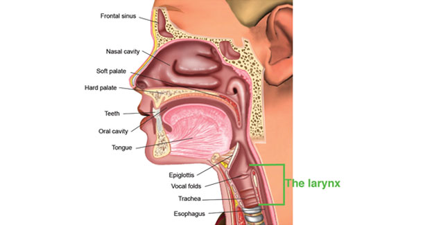
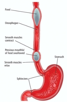
- Oesophagus – is a muscular tube, 25cm long approx., it has a valve at each end called a Sphincter. Its function is to transport food and liquid from the pharynx to the stomach. Food which is now pulp (bolus) is pushed along here by peristaltic action (wavelike contraction of the smooth muscle tissue of the walls). Mucus is secreted by the oesophagus which lubricates the passage of food. No absorption takes place here.
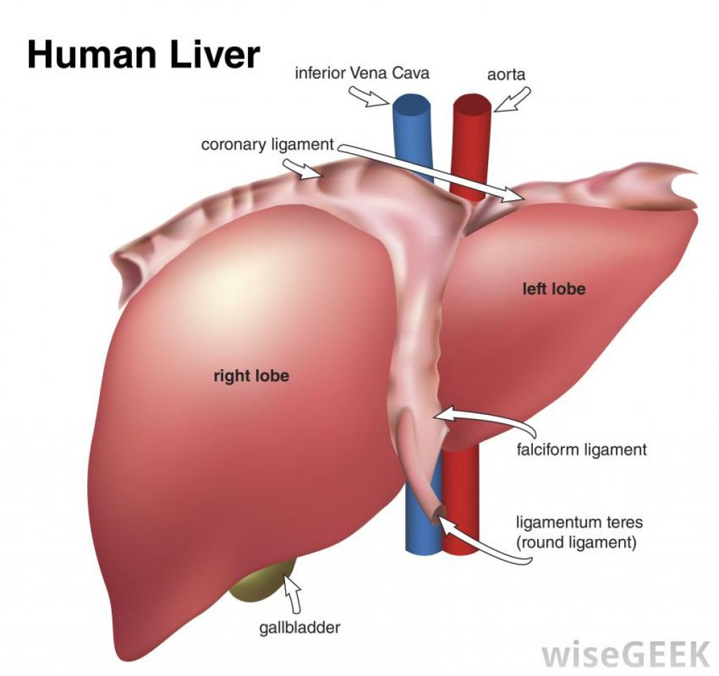
- Liver – is the largest internal organ and largest gland in the body. It weighs approx. 3.5lbs. It has a soft reddish brown colour and is triangular or wedge shaped. It is located under the diaphragm, above the stomach on the right side of the abdomen. The liver has the capacity to regenerate itself.
It has many functions:
Removes
- Harmful substances and toxins from chemicals, drugs and alcohol.
- Nitrogen and Amino Acids
Stores
- Glycogen and Iron
- Vitamins A, B12, D, E, K
Produces
- Heparin – an anti-coagulant
- Bile
- Thrombopoietin – Platelet formation
- Heat – it is the body’s radiator
- Vitamin A – from Carotene (found in green leafy vegetables and carrots)
- Vitamin D – the first stage of Vitamin D synthesis within the body takes place in the liver (Healthy Eating, 2018)
- Plasma Proteins
- Uric acid and urea from the breakdown of red blood cells and amino acids
Converts
- Metabolism of proteins
- Glycogen to glucose when energy is needed
- Glucose back to glycogen when insulin is present
- Stored fats (saturated) into cholesterol
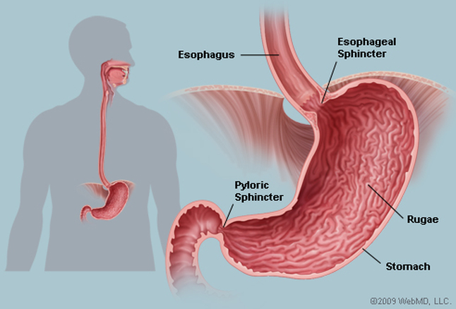
- Stomach – is a C shaped organ, it has layers of muscle fibres that expand and contract. It is lined with a mucus membrane and contains folds called Rugae. It expands when full and contracts when empty.
It is a temporary storage area for food. It churns food (chemical digestion) with gastric juices **. The mucus membrane produces mucus to help lubricate food. It absorbs alcohol directly into the bloodstream. It digests protein through the action of enzymes (chemical digestion). It also forms a thick liquid called Chyme. Chyme is squirted from the stomach into the first part of the small intestine (duodenum).
Gastric glands secretes gastric juices/enzymes as follows: **
| Gastric Glands | Enzymes |
| Hydrochloric Acid – kills bacteria | Pepsin – Breaks down Protein into Polypeptides |
| Sodium Chloride and Potassium Chloride – Assist with the breakdown of Proteins |
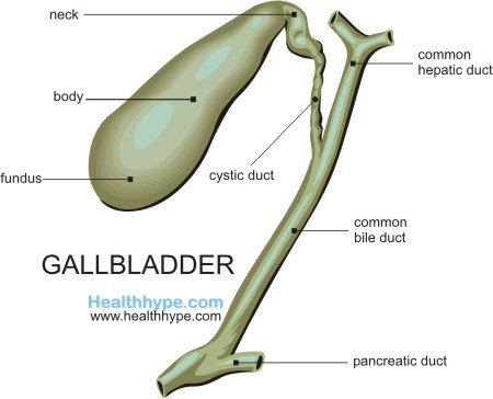
- Gallbladder – is a hollow pear shaped sac attached to the liver by cystic and bile ducts. It’s located under the liver in the upper right hand side of the abdomen. It is a storage area for bile, bile is made in the liver, but is stored in a concentrated form in the gallbladder until needed. It delivers bile to the small intestine. Its lining is made up of many folds, the folds flatten out when it expands. The gallbladder is an ____________ Gland, as it secretes directly into the bloodstream and contains Ducts.
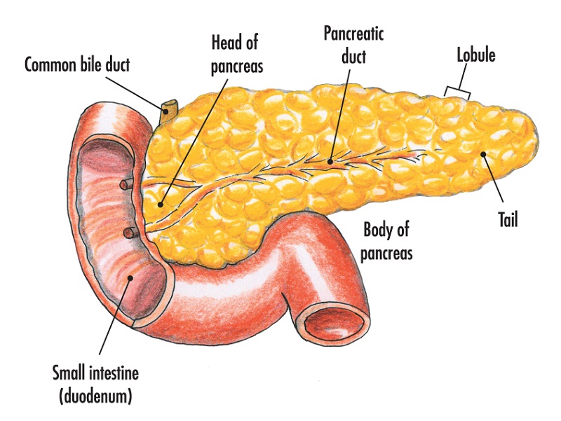
- Pancreas – is a greyish pink long flat gland, is has a fish like shape. It weighs approx. 80g and is 6 inches long. It is located behind the stomach. The head of the pancreas connects to the small intestine (duodenum). It plays a key role in digestion and glucose control. It plays a role in the digestive system and endocrine system. It
It produces very important hormones – Insulin and Glucagon. It is both an Endocrine and Exocrine Gland.
Endocrine – produces hormones
Insulin
Glucagon
Exocrine – produces enzymes
Lipase
Trypsin
Amylase
http://www.innerbody.com/image/endo03.html
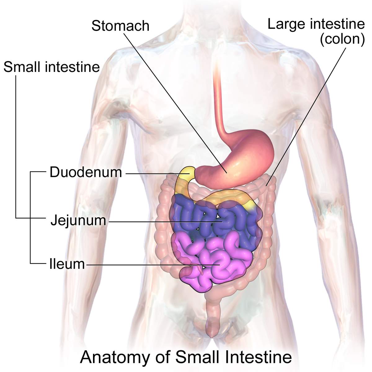
- Small Intestine – is approx. 20 meters long. It is made up of 3 parts, Duodenum (1ft), Jejunum (3-6ft) and ileum (6-12ft). 90% of the digestive process takes place here. The walls (lumen) of the small intestine have many folds called villi and microvilli these tiny finger like projections increase the surface area of the small intestine. They contract and relax, and sway in order to come into contact with most of the contents of the small intestine. All food is moved through the small intestine by Peristalsis – Involuntary wave like contractions of smooth muscle of hollow organs which move food through the body.
Duodenum – C shaped hollow organ located below the stomach and curves around the pancreas. It is the first part of the small intestine and the shortest. It connects the stomach to the Jejunum. It continues the process of digestion which began in the stomach. Its primary function is to receive Chyme a combination of partly digested food and stomach acids into the Duodenum. Mucus is also produced in the folds between the villi in glands called Crypts of Lieberkuhn, the mucus protects the duodenum from the acidic content of chyme. The duodenum also receives bile from the Liver and Pancreatic Juices from the pancreas to aid digestion of food. Bile emulsifies fats and aids in their digestion. Pancreatic juices work as follows: Trypsin breaks down Protein into Amino Acids, Pancreatic Amylase breaks down Carbohydrates into Monosaccharides and Saccharides, Lipase breaks down Fats into Fatty Acids. The spaces in between the villi in the duodenum
Jejunum – is a continuation of the small intestine from the Duodenum. It is the middle segment found between the duodenum and Ileum. The villi in the jejunum are tall and cylindrical. It is located in the upper left hand side of the body. It has a thick intestinal wall. It has longer straight arteries. It is red in colour. This is where the chemical digestion food is completed.
Ileum – is the final part of the small intestine and opens to the large intestine. The villi in the ileum are short and cylindrical. The ileum is located in the lower left hand side of the body. It has a thin intestinal wall. It has shorter straight arteries. It is pink in colour. The main function of the ileum is to absorb nutrients. Bile is also absorbed here, through the blood vessels in the intestinal walls it is returned to the liver. Peyers Patches
http://teachmeanatomy.info/abdomen/gi-tract/small-intestine/
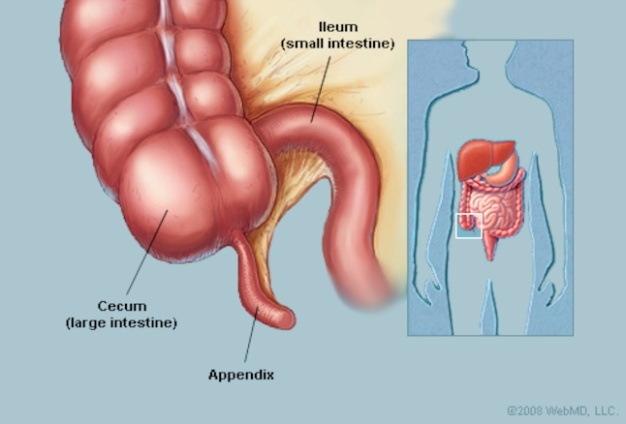
- Large Intestine – it is the last part of the gastrointestinal tract. It is 1.5meters in length. It begins on the right side of the abdomen and is draped around the small intestine. It is shorter than the small intestine but it is thicker in diameter. It is also known as the Colon or Large Bowel. The large intestine is made up of the Caecum, Appendix, Colon, Rectum, Anal Canal and Anus.
The functions of the large intestine are:
- Absorption of nutrients, salt, water and vitamins that remain in digestive waste.
- Secretion – secretes mucus which helps lubricate the passage of waste.
- Produces Vitamin – produces Vitamin K, this plays an important role in blood clotting.
- Breaks down undigested food using bacteria.
- Formation of Faeces – the undigested residue of food is formed here as Faeces.
- Storage of Faeces – the rectum is a temporary storage area for Faeces.
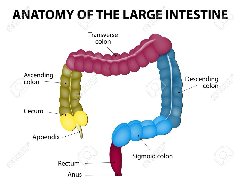
- Appendix – it is located in the lower right hand of the abdomen. It is approx. 4inches long and has a diameter of about ¼inch. It is a narrow pouch of tissue with a wormlike appearance. It consists of a tissue call Lymphoid Tissue which plays a role in the immune system. It is not a vital organ. Scientists are still debating as to what the exact function of the Appendix are. Appendicitis occurs as a result of inflammation of the appendix.
- One theory is that it was used in the past by our ancestors to digest cellulose material from plant based products, which we no longer have a need for or use.
- Another theory is that the appendix is used for or acts as a storage area for bacteria which are beneficial to our bodies during times of illness. These bacteria survive and rebalance our intestines once the illness has left our system.
http://www.innerbody.com/image/dige03.html
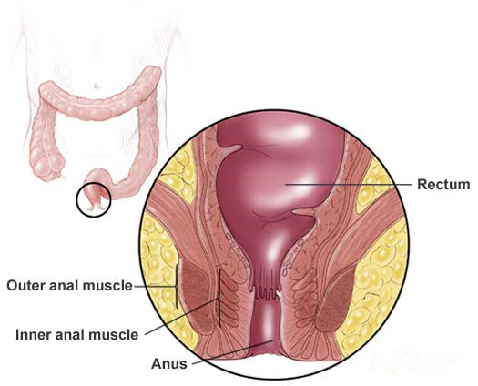
- Rectum – is a short straight section of the gastrointestinal tract. It is approx. 12cm long. This leads from the Colon to the Anus. It is a temporary storage area for faeces. It is usually empty and only receives the contents of the colon when faeces are ready to be expelled from the body from the Anus. Faeces are waste products from food combined with indigestible residues, dead blood cells, fatty acids, mucus and bacteria that are both living and dead. Faeces gets its colour from the pigment in bile called Bilirubin and from dead blood cells.
- Anus – it is the final part of the large intestine, it begins at the bottom of the rectum. The anus is separated from the rectum by the anorectal line. It is the external opening where faeces are expelled/excreted from the body. The anus is surrounded by touch tissues called fascia. Fluid is released into the anus to keep the surface moist. The anus is held closed by the internal sphincter which is under involuntary control. The external sphincter is under voluntary control, this is the only part of the bowel that we have conscious control over.
(iii) Macronutrients
Proteins, Fats and Carbohydrates are known as macronutrients. They provide energy for the body in bulk amounts. Carbohydrates and protein provide 4 calories per 1 gram while Fat provides 9 calories per 1 gram.
| Proteins | Fats | Carbohydrates | |
| Composition | |||
| Digestion | |||
| Absorption |
PART B
(i) Animal Cell
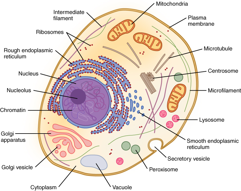
Cells
Cells are the smallest living unit in the body, they are also the building blocks of the body. They are eukaryotic cells, this means that they have a true nucleus and have specialised structures called organelles. Organelles work together to allow the cell to function, they a variety of functions (each with their own function) to perform such as enzyme and hormone production and also providing energy for the cell.
Cells relationship to the body
- A group of cells of similar type join to form tissue
- A group of tissues of related function join to form an organ (heat, lung, stomach)
- A group of organs of related function join to form a system (respiratory, digestive, vascular)
- A group of systems join together to form an organism – the human body
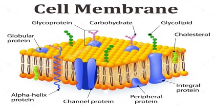
| Organelle | Function | Diagram |
| Cell Membrane |
waste – OUT] |
 |
| Cytoplasm |
|
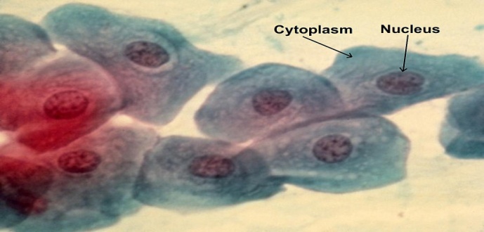 |
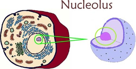
| Nucleus |
|
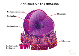 |
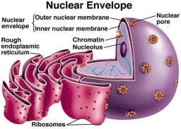
| Nuclear Membrane
and Nucleopore |
|
 |
| Nucleolus |
|
 |
| Mitochondria |
|
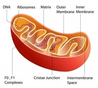 |
| Golgi Apparatus |
|
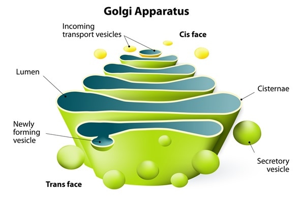 |
| Ribosomes |
|
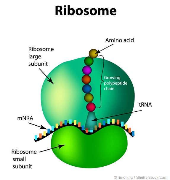 |
| Lysosomes |
|
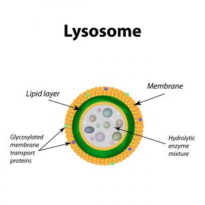 |
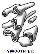
| Smooth Endoplastic Reticulum |
|
 |
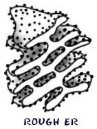
| Rough Endoplastic Reticulum |
|
 |
| Vacuoles |
|
 |
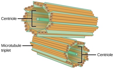
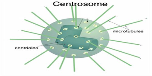
| Centrioles |
|
 |
| Centrosome |
|
 |
(ii) Tissues
Tissue
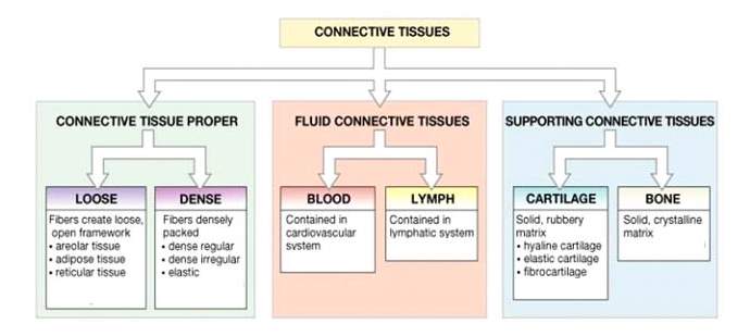
Tissues are groups of cells that are similar in structure and function which act together to form tissues. The study of tissue is known as “Histology”. There are four basic types of tissue in the human body:
- Epithelial Tissue
- Connective Tissue
- Muscle Tissue
- Nerve Tissue
Epithelial Tissue
Epithelia tissue is comprised of epithelial cells. They are widespread in the body. They are closely packed cells, arranged in continuous flat sheets. They have one free surface that is not in contact with other cells. They are avascular – have no blood supply but have an excellent nerve supply – innervated. They cover body surfaces and line hollow organs and the main tissue of glands. They have the ability to regenerate themselves, this means that they can renew themselves. There are 8 types of Epithelial Tissue: 6 of them are identified based on their number of layers and shape, 2 of them are identified by the cell type (squamous) that are found in them. They are usually described with two names, the first name refers to number of layers of cells and the second refers to the cell shape.
Functions of Epithelial Tissue
- Protection – from external factors such as toxins, also protects against physical trauma.
- Absorption – food/substances in the lining of the digestive system.
- Secretion – of enzymes, mucus, sweat and any other substances that are delivered by ducts. Also secretes hormones into the vascular system.
- Detection – of sensations such as touch, cold, heat etc.
- Regulation and Excretion – of chemicals between tissues and body cavities.
| Epithelial Tissue | Example | Diagram |
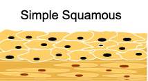
| Simple Squamous Epithelium | Structure: Single layer of flat cells attached to a base membrane.
Function: forms a thin, permeable lining to allow diffusion and filtration for the Alveoli of the lungs, the heart, the blood and lymph vessels. |
 |
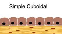
| Simple Cuboidal Epithelium | Structure: Single layer of cub shaped cells attached to a base membrane.
Function: they form the lining of tubules in the kidney and some glands. |
 |
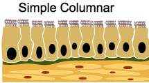
| Simple Columnar Epithelium | Structure: single layer of tall, rectangular cells attached to a base membrane. They are resilient.
Function: form the lining of very active parts of body, such as the stomach, intestines and urethra. Some cells secrete mucus and enzymes, some absorb mucus depending on their position in the body. |
 |
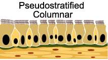
| Pseudostratified Columnar Epithelium | Structure: Falsely stratified single layer of cells attached to a base membrane. The cells appear stratified as they differ in height. All the cells rest on the base membrane however, all of them do not reach the apical surface or surface of the lumen. The nuclei are staggered giving the appearance of stratification.
Function: secretes and move mucus, can be subdivided as follows;
|
 |
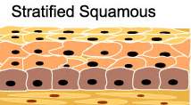
| Stratified Squamous Epithelium | Structure: has two or more layers. This tissue is constantly renewing itself. The bottom layer of cells divide and the new identical daughter cells move towards the surface there they mature and then rejuvenate.
This tissue can be subdivided as follows:
Function: the main function of this tissue is for protection, the higher the number of layers it has the more protection it gives. It is also very good at tolerating abrasion. |
 |
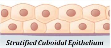
| Stratified Cuboidal Epithelium | Structure: made up of two or more layers. Usually no more than four layers. Round, square or hexagonal in shape. Have equal dimensions on every side. There is a round nucleus in each cell. The bottom row of cells sit on a base membrane. The upper row of cells are freely exposed to a body cavity.
Function: Protection – can withstand chemical and physical assault. Areas that are exposed to higher potential of being damaged such as the oesophagus, vagina, anal canal and most ducts. |
 |
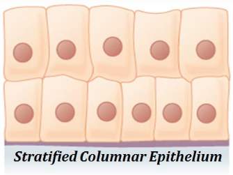
| Stratified Columnar Epithelium | Structure: column shaped cells arranged in multiple layers. They are rare in the body. It mainly occurs in embryonic tissue and not in adults. Can be found in certain glands and ducts but are uncommon in the body
Function: Secretion and protection. Found in the urethra, uterus, vas deferens, eye (conjunctiva), pharynx, salivary glands, anus and lobar ducts. |
 |
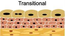
| Transitional Epithelium | Structure: It is only found in the Urinary System – ureters and bladder. It is so called as it gradually changes shape as the bladder fills up with urine. When empty the bladder’s epithelium is convoluted with a convex umbrella shaped surface. When it fills the convolutions are lost and the cells change from cuboidal to squamous. When the bladder is empty the epithelium appears thicker and multi layered. When the bladder is full it is less stratified and more stretched out.
Function: Allows urinary organs to expand and contract. |
 |
Connective Tissue
As the name suggests, connective tissue connects all other tissue in the body. It is widely distributed and the most abundant in the body. It varies in blood supply. Cartilage lacks blood vessels – avascular, dense connective tissue has a poor blood supply. Bone has a rich supply of blood vessels.
It is comprised of three main components: fibres, cells and ground substance. Ground substance is a colourless, clear, gelatinous fluid that fills the space between cells and fibres. The fibres combined with the ground substance make up extracellular matrix. The extracellular matrix provides support to the cells, it also provides biochemical support – regulates the cells dynamic behaviour. Connective tissue is classified into two subcategories: soft and specialised connective tissue.
There are three types of fibres found in connective tissue:
Collagen – fibrous proteins that provide high malleable strength
Elastin – long, thin fibres that allow the tissue to stretch and recoil
Reticular – short, fine collagenous fibres which branch into tiny delicate networks
Types of Connective Tissue
Connective tissue is divided into main categories as follows:
Solid Bone
Semi Solid Fat/Cartilage
Liquid Blood
The Functions of Connective Tissue
- Protection – against impact, friction and contaminants.
- Insulation – thermal insulation (fat)
- Support – structural support (tendons, ligaments, cartilage)
- Storage – stores fuel reserve.
- Transportation – of substances within the body: water and oxygen, vitamins and minerals, energy.
| Connective Tissue | Example | Diagram |
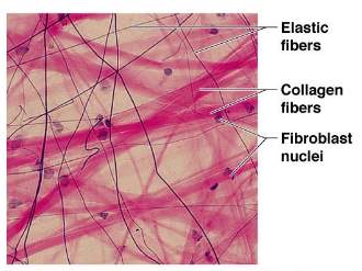
| Loose Connective Tissue:
|
Structure: loose random fibres of loos connective tissue. Wide variety of cell types. Semi Permeable allowing substances to pass through. Contains – yellow and white fibres, fibrocytes, masts cells which produce histamine (protection) and heparin (anti-coagulant, prevents clotting).
Function: Supports and binds other tissues. Defends against infection – when a region of the body is inflamed this tissue soaks up all the excess fluid in the area, this condition is called Edema. Location: Found under the skin, between muscles and in the outer coverings of blood vessels, nerves, oesophagus etc. |
 |
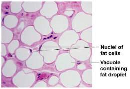
| Loose Connective Tissue:
|
Structure: loose connective tissue, also known as fatty tissue. Consists of fat calls containing fat globules. There are two types (1) white adipose tissue (2) brown adipose tissue: found in specific locations these areas are referred to as adipose depots.
Function: main function is to store energy. Protects and insulates the body. Retains heat as it is a poor conductor of heat. Location: Found under the skin, around the heart, kidneys, eyes, breast and abdominal membranes. |
 |
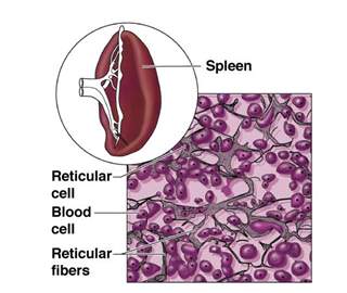
| Loose Connective Tissue:
|
Structure: is a loose connective tissue. It is mesh like. The reticular cells form reticular fibres they form a network in which other cells attach. If forms the scaffolding/framework for soft organs.
Location: Found in the spleen, liver and lymphatic tissue. |
 |
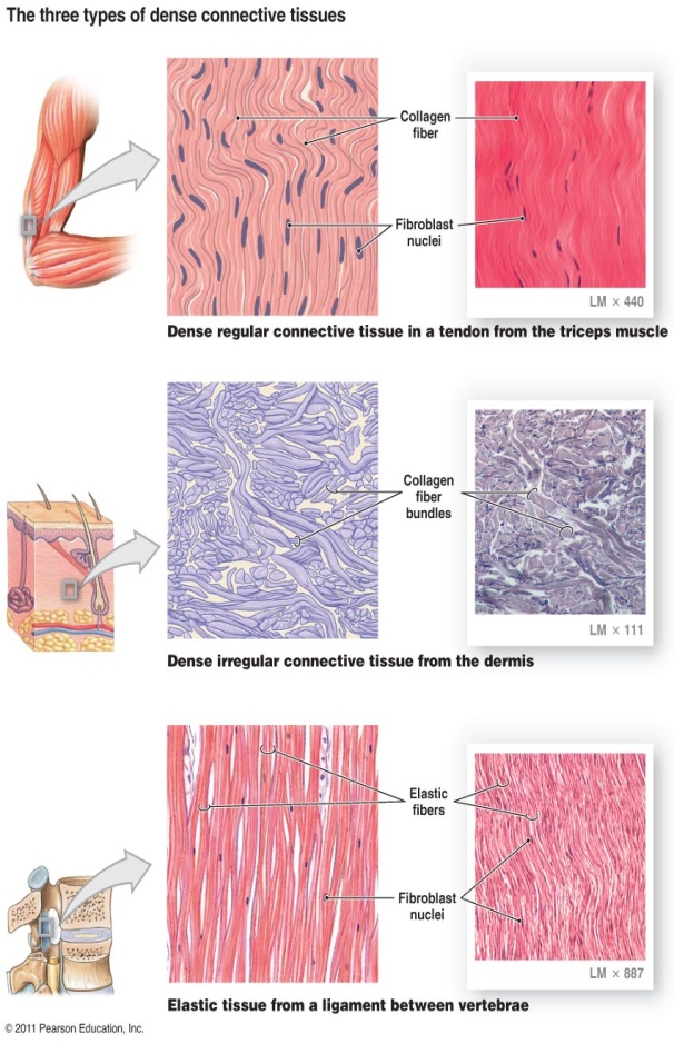
| Dense Connective Tissue:
|
Dense connective tissue contains more collagen fibres than loose connective tissue. As a result it has greater resistance to stretching. Dense connective tissue can be classified as follows:
Function: they enhance tensile strength. Found in ligament and tendons. Also found in the fibrous membranes of blood vessels, nerves and wrapped around muscles.
Function: gives tissue greater strength in all directions and then less strength in one particular direction. Found in the dermis of the skin. Dense irregular elastic tissue in the lungs and arteries give them the ability to regain their shape after stretching.
Function: it allows tissue to recoil, return to its original length after being stretched. Found in walls of bronchial tubes and arterial blood vessels.
|
 |
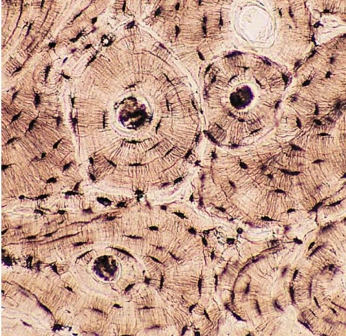
| Bone | Structure: hardest structure in the body. Also called Osseous tissue.
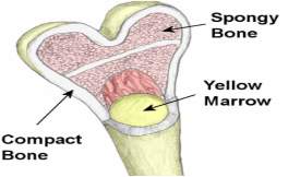
There are two types (1) compact (2) cancellous. Compact is dense bone, it has minimal gaps and spaces. Its porosity is 5–30%. It accounts for 80% of the skeleton. Cancellous is spongy and composed of 25% water, 30% organic material, 45% inorganic salt. Function: Support and protect the body and all the organs. Formation of red blood cells in the bone marrow. Stores and releases phosphorus and calcium. Location: Found in the skeleton. |
|
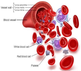
| Blood | Structure: this is a specialised form of connective tissue. It is atypical of this tissue as it does not connect, bind or network with any body cells. It is made up of blood cells – 45% cells and 55% plasma.
Function: transports nutrients, oxygen, hormones, gases and metabolic waste products. Location: Found in Cardiovascular System. |
  |
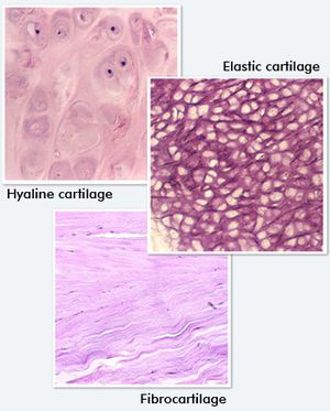
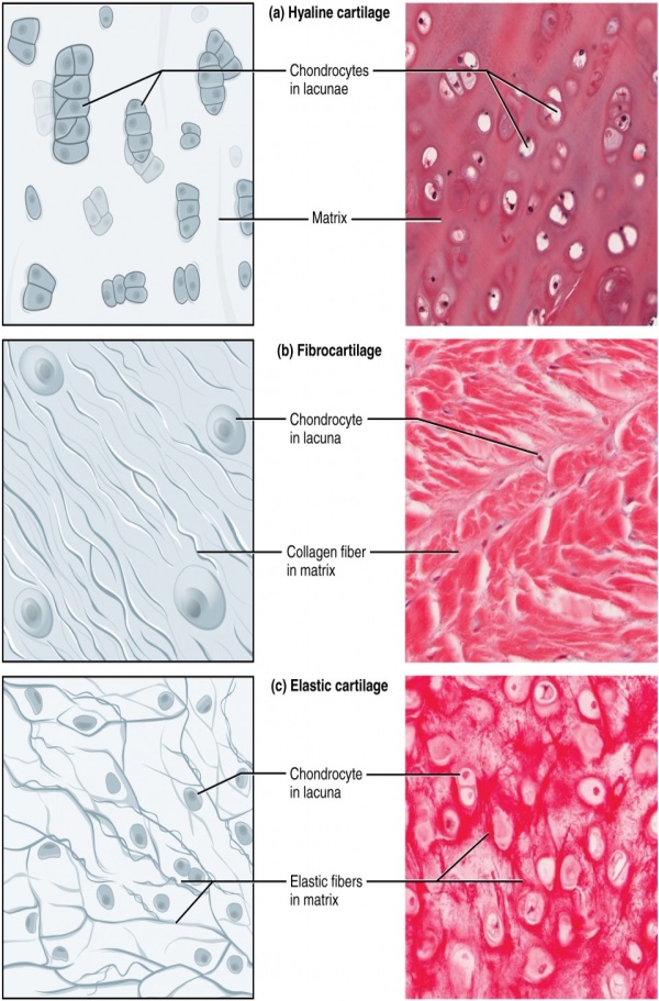
| Cartilage Tissue
cartilage |
Cartilage Tissue: firm, solid, tough, flexible connective tissue. Composed of specialised calls, called Chondrocytes. Cartilage does not contain blood vessels.
Cartilages is classified into three types:
Function: provides cartilage pads for shock absorption, provides support. Found in rib cage, trachea, larynx and nose.
Function: to maintain shape of a structure while still allowing flexibility. Found in the epiglottis and the outer ear – the visible part pinna/auricle.
Function: acts as a shock absorber. Found in joints between bones, found in knee, hip and shoulder sockets. Forms intervertebral discs. |
|
https://courses.lumenlearning.com/nemcc-ap/chapter/connective-tissue-supports-and-protects/
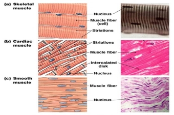
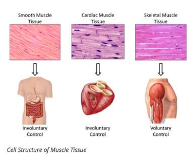
Muscle Tissue
Structure: All muscle cells are made up of 75% water, 20% protein, 5% mineral salts, glycogen, glucose and fat. They are long and fibrous. They are arranged in bundles and parallel lines which makes them very strong. They are likened to a pile of rubber bands that are lines up next to each other, if you attempt to stretch them you will get the idea of what their nature is. They are striated and non-striated. Striated means a striped appearance. Muscle tissue has a striped appearance due to the alternating bands of dark and light protein fibres, Myosin (dark) and Actin (light).
Functions of Muscle Tissue
The main physiology of muscle tissue is movement.
| Muscle Type | Structure and Function | Example |
| Skeletal |
|
Attached to the Skeleton. Found in Biceps and Triceps. |
| Smooth |
|
Found in tissue systems – digestive system and respiratory system. Also found in the walls of hollow organs. Located in oesophagus, stomach, airway, bladder, uterus, blood vessels. |
| Cardiac |
|
Found in the heart only. |
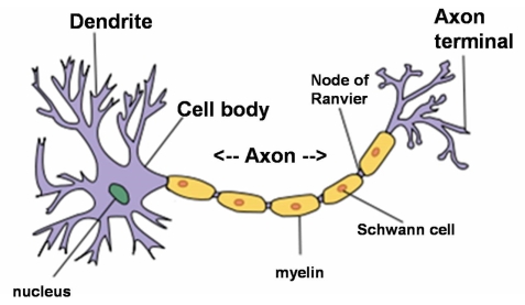
Nerve Tissue
Nerve tissue is found in the Brain, Spinal Cord and Nerves. Nerve tissue is composed of nerve cells called neurons. These are specialised unique cells. They are supported by cells called neuroglia, there are six types of neuroglia. Four types of neuroglia are located in the central nervous system and the remaining two are found in the peripheral nervous system.
Neurons are found in the Central Nervous System and Peripheral Nervous System. The function of neurons is to send and receive nerve impulses in the body.
- Sensory Neurons – send impulses from the body and environment to the brain via the spinal cord.
- Motor Neurons – send impulses from the brain to the body via the spinal cord.
Typical neurons (motor neuron) have cell bodies, dendrites and axons.
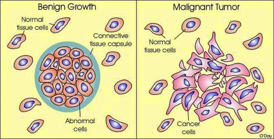
(iii) Tumours
Tumours
Throughout the course of our lives, healthy cells in our bodies, multiply, divide and renew themselves in a controlled way. A tumour also called a Neoplasm occurs when cells escape from the normal process of cell division, they multiply at an abnormal rate forming a mass or a lump. Tumours may be Benign or Malignant. The two most important differences between Benign and Malignant are invasion and spread.
Benign Tumour
A Benign Tumour is an abnormal growth that serves no purpose. It is non-cancerous. It can cause pressure or pain in the area affected. It is enclosed in a capsule. It does not invade or spread and usually does not re-occur.
Causes of Benign Tumours
The cause is often unknown, but may be linked to
- Genetics
- Diet
- Stress
- Inflammation or infection
- Localised trauma or injury
- Environmental toxins or exposure to radiation
Types of Benign Tumours
Adenomas – they start in the epithelial tissue of glands, also may be found in pituitary, thyroid and adrenal gland, and may be found in the liver. Can be removed with surgery.
Fibromas – fibrous tissue or connective tissue tumours. They can form in an organ, most common in uterus. They are symptom causing (vaginal bleeding, pelvic pain and bladder issues) and need to be removed surgically.
Hemangiomas – this is an accumulation of blood vessel cells in the internal organs or on the skin (birthmark). They are red or bluish in colour and those that interfere with sensory organs i.e. hearing, vision can be treated with medication – corticosteroids.
Lipomas – develop in fat cells, these are the most common benign tumours that are found in adults. They are slow growing and found in arms, neck, back and shoulders. Can be surgically removed if becomes painful.
Meningiomas – these benign tumours develop in the membrane of the brain and spinal cord. Slow growing. Variety of treatment – ones that can’t be surgically removed are treated with radiation, surgical treatment depends on location in the body, age, and whether it is attached. Symptoms include visual problems, seizure, personality changes, headaches and weakness on one side.
Myomas – tumours that develop from muscle. Skeletal muscle tumour is called Rhabdomyoma. Smooth muscle tumour found in internal organs (uterus and stomach) are called Leiomyomas. They may be surgically removed or shrunk with medication.
Nevi – growths that develop on the skin. They range in colour and shape and can develop into a melanoma (skin cancer). It is important to have any abnormalities on the skin checked by a health care professional. It may be necessary to remove growths such as moles, warts and have them checked for signs of cancer.
Neuromas – tumours that develop from nerves. There are two types Schwannomas and Neurofibromas (more common in people who have Neurofibromatosis. Can happen anywhere in the body where there are nerves. Benign nerve tumours are surgically removed.
Osteochondromas – benign tumours of the bone. Appear near the joints as a bump, found in the shoulder or knee. Will be surgically removed if causing pressure on nerves or blood vessels causing pain.
Papillomas – finger like projections that develop in epithelial tissue. These tumours can be benign or malignant. Can be found in the breast duct, cervix, skin and mucous membrane in the eyelid. They can go away on their own or can be surgically treated. Usually caused by coming in contact with the human papillomavirus infection.
Malignant Tumour
The term Cancer includes more than 100 diseases, which are all potentially life threatening. It is a human ailment. Malignant tumours are cancerous. They can invade and spread to other organs and tissue, they can also spread through blood and lymph. Seedlings break off and travel to other body parts where they metastasise. They often re-occur.
Causes
The cause have been debated and studied for years amongst the medical community. Causes are thought to be hereditary but also from:
Environmental factors:
- Smoking – lung cancer
- Alcohol – cancer of the liver
- Diet and Exercise
- Chemical Exposure
Symptoms of tumours
- Local Symptoms – pain in the area, lumps, swelling and haemorrhaging.
- Metastatic Symptoms – enlarged spleen (splenomegaly), enlarged liver (hepatomegaly), enlarged lymph nodes.
- Systemic Symptoms – symptoms are more generic weight loss, fatigue, sweating, anaemia etc.
Types of Malignant Tumours
Carcinomas – most common cancer, develop in lungs, skin, pancreas, breasts and other glands.
Sarcomas – relatively uncommon form of cancer, found in blood vessels, bone, fat, cartilage and other connective tissues in the body.
Melanomas – type of skin cancer, caused by mutations of DNA in melanocytes (cells). Good prognosis if detected early e.g. mole or wart
Lymphomas – this cancer develops in the lymphatic system, it is a cancer of the lymphocytes (white blood cells) it can develop anywhere in the body. They behave in an abnormal manner and are unable to fight infection, they collect in the lymph nodes, the spleen and bone marrow.
Leukemia – cancer of the blood, usually does not form a solid tumour. It is whereby the bone marrow makes too many lymphocytes (white blood cells), abnormal cells cause a build-up in the blood. Leukemia is more common in children.
Treatment
Treatment is determined by location, size and the stage of progression of the tumour. Treatment methods are Radiation, Chemotherapy, Hormone Therapy and Surgery. Surgery is mostly offered to patients to remove the tumour and prevent reoccurrence. Prognosis is good if detected early.
The Urinary System
The urinary system or the renal system is the organs, muscles, tubes and nerves that work together to produce, store and transport urine. The main function of the urinary system is to remove excess metabolic waste products and excess fluid from the body. The urinary system consists of the kidneys, ureters, bladder and urethra, collectively known as the urinary tract, this is the pathway that urine is transported through. It produces a hormone that controls the rate of red blood cell formation. It produces an enzyme that controls blood pressure. These functions are essential for homeostasis – the ability to maintain an internal body environment.
Kidney
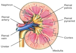
Structure of Kidney
The human body has two kidneys. They are dark red in colour. They are bean shaped organs. They are 11cm long and 6cm wide. They are located towards the back of the abdominal cavity, just above waist level. Each kidney is enclosed in a transparent membrane called the renal capsule, this helps to protect against trauma and infection. There are three main areas of the kidney: Renal Cortex, Renal Medulla and Renal Pelvis.
Renal Cortex – the renal cortex is the outer part of the kidney. It has smooth texture and is reddish in colour. It is the location of the Bowman’s Capsule and the glomeruli, in addition to the proximal and distal convoluted tubules and their associated blood supplies (these structures are part of the nephrons).
Renal Medulla – the renal medulla is the inner part of the kidney. Medulla meaning inner portion. This area is red-brown in colour and is striated (striped).
Renal Pelvis – collection area for urine. The renal pelvis is a funnel shaped cavity that receives the urine that has been drained from the nephrons in the kidney via the collecting ducts and then the papillary ducts (larger ducts).
Renal Hilus – the renal hilus is an indentation near to the centre of the concave area of the kidney. This is the area of the kidney through which the ureter leave the kidney together with other structures which include blood and lymphatic vessels and nerves that enter and leave the kidney. It the entry point to the kidney.
Renal Capsule – the renal capsule is a smooth, transparent, fibrous membrane which surrounds, protects and encloses the kidney. Each kidney has its own renal capsule which helps maintain the shape of the kidney and also protects it from damage. The renal capsule itself is surrounded by a mass of fatty tissue which also protects the kidney from damage by cushioning it in case of impact or sudden movements.
Renal Pyramids – within the renal medulla of each kidney there are approx. 5-18 striated triangular structures. The striations appear due to the many straight tubule and blood vessels within the renal pyramids.
Rental Artery – the renal artery delivers oxygenated blood to the kidney. This is a main artery and divided into many smaller branches as it enters the kidney via the renal hilus. These eventually separate into afferent arterioles (bring blood to the glomerulus) one of which serves each nephron in the kidney.
Renal Vein – the renal vein receives deoxygenated blood from the veins within the kidney, through which it is returned to the blood circulation system.
Interlobular Artery – the interlobular artery delivers blood at high pressure to the glomerular capillaries.
Kidney Nephron – kidney nephrons are the functional units of the kidneys. It is the kidney nephrons that actually perform the main functions of the kidneys. There are approx. a million nephrons contained within each kidney.
Collecting Duct (kidney) – the collecting duct is part of the kidney nephron. Many of these such collecting ducts unity to drain urine that is extracted by the kidney.
Function of Kidney
- Filters Blood – removes waste and excess water from blood.
- Formation of Urine – separates waste and water from filtrate, what is not reabsorbed is urine.
- Production of hormones
- Renin – regulates sodium levels
- Erythropoeitin – stimulates production of red blood cells.
- Regulates Blood Pressure – produces an enzyme called Angiotensin
- Produces Vitamin D
Ureters
Structure of Ureters
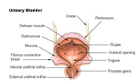
The ureters are two slender, muscular tubes which carry urine from the kidneys to the bladder. The ureters are wider at the top where they come into contact with the renal pelvis, but they quickly become very narrow. Where the two ureters enter the bladder, a small triangular fold of tissue call the trigone keeps urine from flowing back into the ureters, after it has emptied into the bladder. They are lined with a mucous membrane which helps protect against infection.
Function of Ureters
The involuntary wavelike contraction of the smooth muscle in the ureters moves urine away from the kidney and towards the bladder. This is known as peristalsis.
Bladder
Structure of Bladder
The bladder is a hollow muscular sac like organ that sits on the pelvic floor. It is made from transitional epithelium tissue. It is holding place for urine. The walls of the bladder is lined with a mucous membrane, it has three layers of smooth muscle. Urination occurs when the walls of the bladder contract. The internal sphincter keeps the bladder closed while it fills – this is a ring of involuntary muscle located where the bladder and urethra join. The external sphincter relaxes to allow urine to pass during urination – this is a ring of voluntary muscle.
Function of Bladder
The function of the bladder is to store urine that is produced in the kidney and excrete it from the body. A moderately full bladder contains about 470ml of urine, when the urine collected reaches 300-350ml the internal sphincter (involuntary, controlled by the autonomic nervous system) opens, urine floods into the upper part of the urethra. The urge to urinate occurs at this point. The external urethral sphincter is then relaxed voluntarily, the muscles of the bladder contract, allowing urine to pass out of the body through the urethra. Even though it is possible to delay urination for a period of time, the bladder will eventually empty itself automatically. The bladder also has a minor role in regulating temperature as some heat may leave from the body in the form of urine.
Urethra
The urethra is a tube that connects the bladder to the genitals for the excretion of fluids from the body. The external sphincter is made of striated muscle which allows voluntary control of the flow or urine from the bladder into the urethra.
Female Urethra
The urethra in females is straight shaped and relatively shorter than males (1½ – 2½ inches in length). This makes them more susceptible to bacterial UTIs – Urinary Tract Infections. It is located above the opening to the vagina. The lining is primarily composed of stratified squamous epithelium – non keratinised, which becomes transitional as it nears the bladder. It comprises of three layers:
- Smooth muscle tissue – continuation of smooth muscle tissue of the bladder.
- Erectile tissue – specialised tissue that becomes engorged with blood when sexual arousal occurs.
- Mucous membrane tissue – protects the urethral tissues from corrosion of urine.
Male Urethra
The urethra in males is 6 – 8 inches in length. It is an ‘S’ shaped narrow fibromuscular tube. It is a single structure. It has a series of segments: It has a similar structure to the female urethra. It travels through the penis and transport semen as well as urine. Semen which is made up of sperm cells along with other fluids is carried into the urethra through the vas deferens. The urethra in men is divided into three parts:
- Prostatic urethra – crosses the prostate gland
- Membranous urethra – passes the external sphincter
- Spongy urethra – travels the length of the penis
Nephron
Urine Production in the Nephron
- Filtration in the Bowman’s Capsule
- Blood enters the kidneys via the afferent arterioles. In the kidney there are approx. 1 million nephron, these are the tiny units that clean the blood.
- Each nephron has a glomerulus and a series of tubules. The glomerulus is located in a structure known as the Bowman’s Capsule, the glomerulus is a capillary bed.
- The blood in these capillaries are under pressure, the capillary walls are permeable to water and other substances, these pass through into the capsule, blood cells and proteins remain the blood vessels.
- Therefore the Bowman’s Capsule acts as a collection point for the waste products carried within the blood.
- At the point however, the capsule has also collected other substances which are NOT waste, these will be reabsorbed as they pass through the nephron.
- Re-absorption in the Convoluted Tubules
- This liquid known as Filtrate flows into the twisted tubes known as convoluted tubules.
- The tubes that lead away from the Bowman’s Capsule are known as the Proximal Convoluted Tubules.
- These straighten into a long loop called the Loop of Henle, which then passes into the medulla and back to the cortex.
- The final series of twisted tubes is called the Distal Convoluted Tubules.
- As the filtrate flows through the tubules the capillaries reabsorb useful substances such as water, nutrients and minerals back into the bloodstream.
- Only 1% of the liquid into the Bowman’s Capsule is excreted in urine. The rest is reabsorbed.
- Collection in the Pelvic Calyces (Renal Pelvis) and Secretion
- By the time the filtrate reaches the end of the tubules only excess fluid and waste products remain. This is Urine. The kidneys produce 1-1 ½ litres of urine per day.
- The nephron straightens out into a collecting tube in the medulla.
- These collecting tubes form pyramid like masses. These pyramids collect the waste products and funnel it into the renal pelvis/pelvic calyces. This is the collection point for urine. It from here that the urine empties into the ureter.
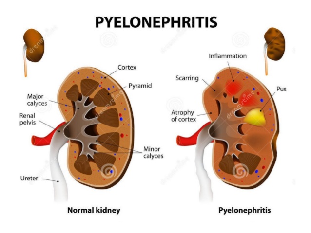
Kidney Infection
Overview
Pyelonephritis or kidney infection is one type of a UTI – Urinary Tract Infection. It usually begins in the urethra or bladder and then travels to one or even both kidneys. It is advisable to respond quickly to the infection as if not treated properly it can cause permanent damage to your kidneys or bacterial can spread to other areas through the bloodstream causing life-threatening illness/infection.
Symptoms
- Fever
- Chills
- Frequent Urination
- Strong urge to urinate which is persistent
- Abdominal, Back, Flank (side) or Groin Pain
- Pain or burning sensation when urinating
- Nausea and Vomiting
- Urine that is cloudy, contains pus or blood, has a bad smell
Causes
The most common cause of kidney infection is when bacteria enter your urinary tract through the urethra, bacteria multiply and travels to the kidneys. Urine is usually sterile, infection arise from bacteria – Escherichia and E. Coli and microorganisms called Chlamydia and Mycoplasma (this may be sexually transmitted). A bacterial infection from elsewhere in the body can also travel through the bloodstream to the kidneys. Complications include: Kidney Scarring, Septicaemia (blood poisoning) and in pregnancy – low birth weight in babies.
Risk Factors
- Being female – females have a shorter urethra than men, shorter passage for bacteria to travel.
- Weakened Immune System – includes medical conditions such as HIV and diabetes.
- Urinary tract blockage – anything (kidney stones) that slow down the flow of urine.
- Nerve damage around the bladder – sensations being blocked by nerve or spinal cord damage.
- Urinary catheter – using a urinary catheter.
- Vesicoureteral reflux (kidney reflux) – where urine in your bladder flow back up into the ureters.
Treatment usually includes antibiotics, in severe cases hospitalisation is required. In the case of structural issues with kidneys, surgery is required.
Prevention
- Drinking fluids – especially water
- Urinate as soon as you need to go!
- Emptying bladder after intercourse
- Avoid the use of feminine products in the genital area
- Use care when wiping – wipe from front to back especially after bowel movements, prevents the spread of bacteria.
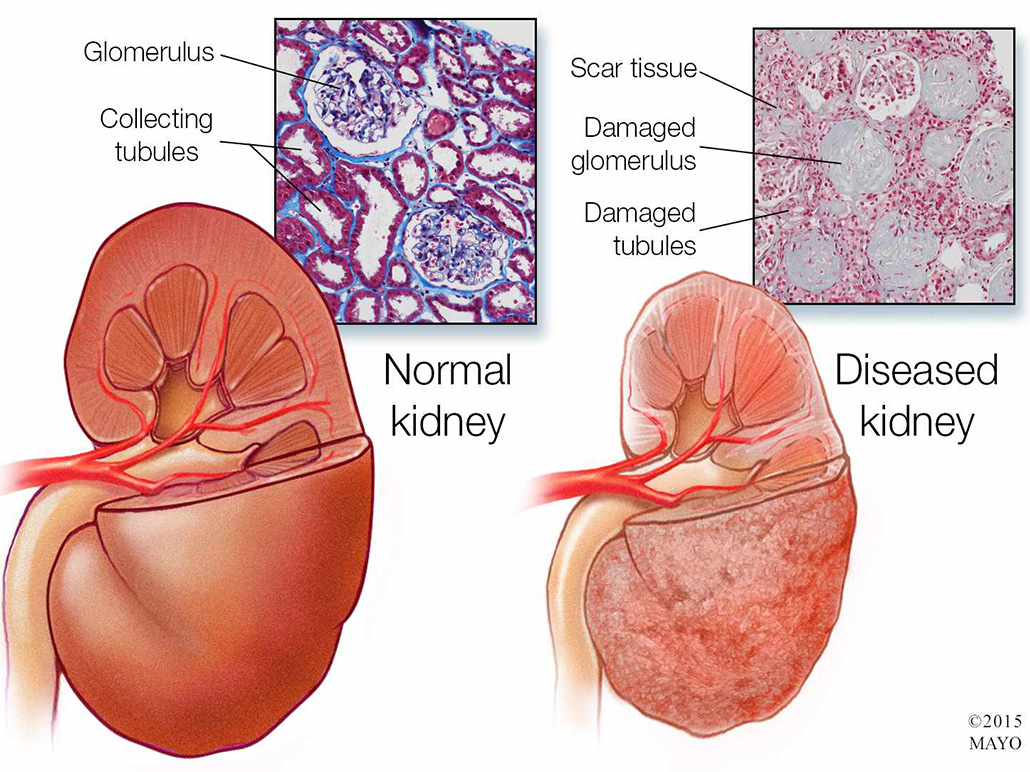
Chronic Kidney Disease
Overview
Chronic kidney disease is also known as chronic kidney failure. It is the gradual loss of kidney function. When the disease reaches an advanced stage dangerous levels of waste, fluid and electrolytes build up in the body. In the early stages symptoms may not be apparent until your kidney function is significantly compromised.
Symptoms – signs and symptoms develop over time and progress slowly, these include:
- Nausea and Vomiting
- Weakness and Fatigue, also mental fatigue
- Loss of appetite
- Changes in urination
- Itching, muscle twitches and cramps
- Shortness of breath – build-up of fluid in lungs
- Chest pain – build-up of fluid in lining of heart
- Swelling of feet and ankles
- Hypertension – high blood pressure
Causes
- Diabetes – type 1 or type 2
- Hypertension – high blood pressure
- Polycystic kidney disease
- Pyelonephritis – recurrent kidney infection
- Prolonged periods of obstruction – kidney stones, enlarged prostate and some cancers
- Vesicoureteral Reflux (kidney reflux) – where urine flows back up into kidneys
- Glomerulonephritis – inflammation of the glomeruli, filtering units of the kidney
- Interstitial Nephritis – inflammation of the tubules and surrounding structures in the kidney
Risk Factors
- High blood pressure
- Diabetes
- Cardiovascular Disease – heart and blood vessels
- Obesity
- Smoking
- Family history of the disease
- Old age
- Abnormality of the kidney structure
- Race – Native American, African American and Asian American origin
Treatment
- Dialysis – artificial removal of waste products and extra fluid from blood when the kidney are no long able to do this.
- Kidney Transplant – surgically placing a healthy kidney from a donor into a person with chronic kidney disease.
Prevention
- Over the counter medication – always follow the instructions carefully, take correct dosage – over use of pain medicine may lead to kidney damage. Consult with doctor and pharmacist on safe drug usage.
- Do not smoke – smoking cigarettes damages your kidneys.
- Maintain a healthy BMI/Weight
- Management of ongoing medical conditions and diseases with healthcare professionals.
Complications
- Fluid retention
- Anaemia
- Pericarditis – inflammation of membrane that surrounds the heart
- Cardiovascular disease – heart and blood vessels
- Pregnancy complications
- End stage kidney disease – irreversible kidney damage
- Hyperkalemia – increase in potassium levels which weakens hearts ability to function
- Damage to central nervous system
- Decrease in immunity – more vulnerable to infection
- Erectile dysfunction, reduced fertility and decreased sex drive
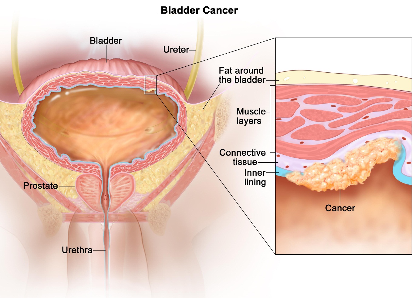
Bladder Cancer
Overview
Bladder cancer is one of the most common cancers, it occurs more frequently in men rather than women. It can happen at any age but mostly affects older adults. Bladder develops usually in urothelial cells which line the inside of the bladder, however, this type of cancer can also develop in other parts of the urinary tract. 7/10 people develop early stages of bladder cancer and is very treatable, however, bladder cancer at this early stage can often reoccur therefore patients require follow up testing for years afterwards.
Types of Bladder Cancer
- Urothelial Carcinoma – previously known as Transitional Cell Carcinoma, this occurs in the cells that line the bladder. These cells expand when the bladder is full and contract when the bladder is empty. These cells are also found in the urethra and the ureters, tumours can form here also.
- Squamous Cell Carcinoma – this is associated with chronic irritation of the bladder most commonly resulting from infection, also from use of urinary catheters.
- Adenocarcinoma – develop in the cells which make up mucus secreting glands in the bladder.
Symptoms
- Hematuria – blood in urine
- Pelvic Pain
- Back Pain
- Painful Urination
- Frequent Urination
Causes – it is not always clear what causes bladder cancer, patients with bladder cancer do not have any obvious risk factors. Bladder cancer develops when bladder cells multiply and grow at an abnormal rate and form lump or a mass called a tumour. Bladder cancer causes include:
- Smoking
- Radiation Exposure – in a work environment
- Exposure to Chemicals – past exposure to radiation
- Chronic irritation of the bladder lining
- Parasitic Infections – in people who travel outside of the United States
Risk Factors
- Smoking – accumulation of harmful chemicals in the urine
- Age – bladder cancer increases as you age, rarely found in patients under the age of 40
- Race – white people are at a greater risk of bladder cancer
- Being Male – men develop bladder cancer more than women
- Chemical Exposure – people who work with chemicals are at greater risk of bladder cancer due to their exposure to chemicals such as arsenic and manufacturing chemicals (rubber, dyes, leather, paint). Kidneys play a vital role in filtering harmful substances from your blood stream.
- Family History – Bladder cancer can reoccur. You can have an increased risk of bladder cancer is your parent, sibling or child have had a history of bladder cancer.
- Chronic Bladder Inflammation – chronic bladder inflammations and urinary infections, long term use of a catheter can lead to bladder cancer
- Cancer Treatments – anti cancer drug treatment cyclophosphamide increases the risk of bladder cancer.
Treatment
- Surgery – removal of cancerous tissue
- Reconstructive Surgery – to create a new exit point in the body for urine when bladder has been removed
- Chemotherapy (intravesical chemotherapy) – treatment of bladder only
- Chemotherapy (systemic chemotherapy) – treatment of the whole body
- Radiation Therapy – where surgery is not an option
- Immunotherapy – to help trigger the immune system in the body to fight the cancer cells
Prevention
- Do Not Smoke – cancer causing chemicals are unable to collect in your bladder.
- Diet – Antioxidants found in fruits and vegetable can help to reduce your risk of cancer.
- Chemical Exposure – follow strict health and safety procedures to avoid exposure.
References
Healthy Eating. (2018). Retrieved from http://healthyeating.sfgate.com/correlation-between-vitamin-d3-liver-10148.html .
www.vivo.colostate.edu/hbooks/pathphys/digestion/liver/metabolic. (2018). Retrieved from Vivo Books.
Cite This Work
To export a reference to this article please select a referencing stye below:
Related Services
View all


DMCA / Removal Request
If you are the original writer of this assignment and no longer wish to have your work published on UKEssays.com then please click the following link to email our support team:
Request essay removal