Functional and Structural Factors for Different Animal Tissues
| ✅ Paper Type: Free Essay | ✅ Subject: Biology |
| ✅ Wordcount: 2180 words | ✅ Published: 23 Sep 2019 |
Construct a table, listing the structure and function of the different plant tissues described in this section (e.g. phloem). Then write a short essay (700 – 1000 words) briefly describing both the functional and structural properties of each of the four animal tissues described in this section.
|
Tissue |
Category |
Location in Plant |
Structure |
Function |
|
Epidermis |
Simple 1 |
Outer surface of all plant organs2 (e.g. leaves.1) |
Upper and lower epidermis made of a single layer of epidermal cells that contain cutin which forms a waxy cuticle on their outward facing surface.2 |
Secretion of a waxy cuticle that prevents loss of water 1, regulating transpiration. 3 |
|
Cork |
Simple 1 |
Roots and stems of older plants. 4 |
Dead cells with thick walls containing lipophilic suberin.4 |
Replaces the stem epidermis in older plants to prevent water loss and to protect the stem and roots from damage.1 |
|
Parenchyma |
Simple 1 |
Most plant organs (e.g. roots, stem and leaves) 1 |
Thin-walled living cells with an unspecialised structure so they can carry out different functions.5 |
Storage of food, tissue renewal and photosynthesis.1 |
|
Collenchyma |
Simple 1 |
Stems, roots1 and leaves. 6 |
Elongated cells with irregularly thick cell walls containing a lot of cellulose. 6 |
Maintaining plant structure 6 and storage of starch.1 |
|
Sclerenchyma |
Simple 1 |
Bark and mature stems (regions that don’t grow) 7 |
Dead, rigid cells due to thick secondary wall containing lignin.1 |
Structural support and protection from damage. 1 |
|
Xylem |
Complex 1 |
Roots and the stem 8 |
Dead, elongated cells in the shape of tubes. Formed from tracheids, xylem vessels, fibers and parenchyma cells.9 |
Where transpiration (transport of water from roots to leaves) occurs. 10 |
|
Phloem |
Complex 1 |
Leaves and stem 11 |
Made up of sieve tubes, companion cells, parenchyma cells and fibres.11 |
Transport of food from leaves throughout the plant.1 |
Figure 1: Table to show the structure and function of different types of plant tissues and where they are located on a plant
Structural and Functional Properties of the Four Types of Animal Tissue
Connective tissue mainly consists of the extracellular matrix1 (University of Kent, 2018), a substance containing proteins and fluids, which provides the tissue with strength and structure12.(Mosby’s Medical Dictionary, 2009) Collagen is an extremely important protein from the extracellular matrix as it has a triple stranded helical structure which makes it very stiff, ideal for giving tissue strength.13 (K. Gelsea, E. PÖschlb, T. Aigner, 2003. Collagens—structure, function, and biosynthesis)

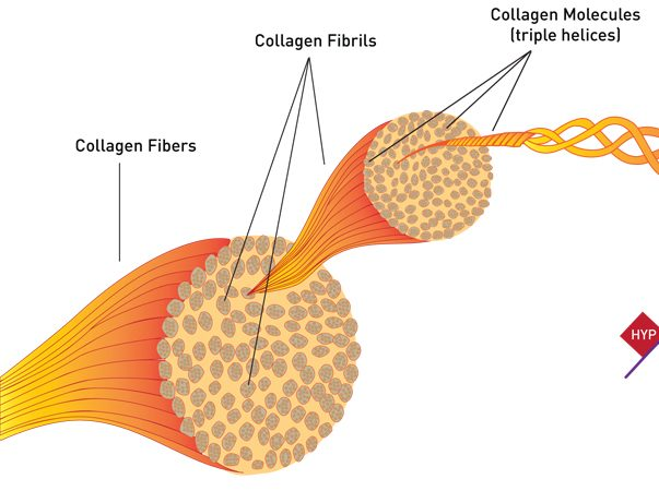
Figure 2: Image showing the triple stranded helical structure that makes up the collagen fibers.14 (Lina Jakimaviciene, 2018. What to do to have perfectly healthy hair?)
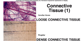
Figure 3: Image showing the structural difference between loose and dense connective tissue. Dense tissue is much firmer whereas loose tissue allows for movement of joints.15 (Dr. Ray Burkett, 2006. A&P I – Tissue Lab)
Epithelial tissue also relies on connective tissue for support, as the basal lamina is made of extracellular matrix arranged into a tough sheet.1 (University of Kent, 2018) The tissue is composed of epithelial cells and desmosomes which form continuous sheets that cover organ walls, line internal cavities and surround glands. Molecules are prevented from leaking across the epithelium by tight junctions within the sheets, helping the epithelium tissue contribute towards its role as a barrier to regulate transport of substances into and out of a cell. The apical surface of epithelial tissue is on the outside of the cell and is exposed to fluids containing nutrients that are to be absorbed into the cell. Epithelium cells of the small intestine have microvilli on their apical surface which increases the surface area, therefore increasing the rate of absorption.1 (University of Kent, 2018) Some cells have cilia on their surface, for example the fallopian tubes have ciliated epithelial cells to help the egg cells travel towards the uterus.16(Study.com, n.d. Ciliated Epithelium: Function, Structure & Diagram)
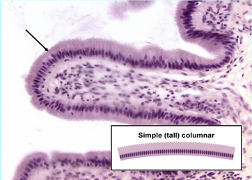
Figure 4: Image of columnar epithelial tissue with an arrow pointing to the microvilli that increase absorption rate 17 (SOCRATIC Anatomy and Physiology, 2016. What is the function of the simple columnar epithelium?)
There are three different forms that epithelial cells can take: squamous, columnar and cuboidal. Squamous cells form one cell thick (simple) epithelial layers in blood capillaries that allow gases (oxygen and carbon dioxide) to easily diffuse in and out of the blood at the lungs and muscles.1 (University of Kent, 2018) As shown by Figure 4, columnar cells are very absorptive due the microvilli on their surface.17 (SOCRATIC Anatomy and Physiology,2016. What is the function of the simple columnar epithelium?) Cuboidal cells line small cavities in the body and areas specialised for secretion and can withstand more pressure due to their sturdier structure.18 (The Virtual Biology Labs, n.d. Simple cuboidal epithelium) Stratified epithelium tissue consists of more than one layer of cells and is protective as it can withstand high abrasion, making it ideal to line places such as the oesophagus.19 (Histology Guide, University of Leeds, 2003. Epithelium: Stratified Epithelia)
Another vital animal tissue is nervous tissue, which transmits nerve impulses around the body to allow movement of muscle and trigger release of hormones. Nervous tissue is made up of neurons (conducting cells) that transmit signals, and neuroglia (non- conducting cells) that protect the neurones.1 (University of Kent, 2018)
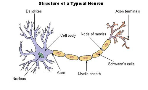
Figure 5: Labelled diagram showing the generic structure of a neuron 20 (Alan Barker, 2009. Learning to juggle helps your brain grow – official!)

Figure 6: Diagram showing the difference in structure between the three types of neurons. Sensory neurons receive signals from receptor cells and motor neurons are connected to effectors. 22 (Joseph Sparks, n.d. Biopsychology: Sensory, Relay and Motor Neurons)
Electrical impulses sent along the central nervous system can reach voluntary muscle tissue which then moves according to a conscious thought. Microfilaments containing the contractile proteins actin and myosin work together and use ATP to move muscle fibres.
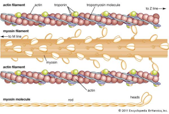
Figure 7: Diagram showing the structure of myosin and actin, and how they interact with one another (myosin head binds to troponin on an actin filament)24 (Encyclopaedia Britannica, n.d. Myosin)
There are three main types of muscle cells: cardiac, smooth muscle and skeletal. Cardiac muscle cells are located in the heart and work together to synchronise the heartbeat with the aid of intercalated disks that join the cells to one another.1 (University of Kent, 2018) Electrical impulses are carried through the heart via the myocardium, a layer of muscle fibre in the heart wall that can conduct electricity, triggering the atria walls contract and push blood into the ventricles. The electrical activity travels to the apex of the heart and up through Purkyne tissue (thin muscle fibres) resulting in strong, continuous contractions.25 (CGP, 2016. AQA A-Level Biology) This is an example of involuntary muscle tissue controlled by the autonomic nervous system, where there is no conscious thought controlling the movement. Smooth muscle, situated in blood vessels and hollow organs also contracts involuntarily, but slower and weaker. Skeletal muscle is striated body muscle connected to bone by tendons that aids voluntary movements controlled by the peripheral nervous system, resulting in a fast and strong discontinuous reaction.1(University of Kent, 2018)
- University of Kent, 2018. Cell Biology: Cells to Systems Study Guide. [Online] Available from: https://moodle.kent.ac.uk/2018/mod/book/view.php?id=237532 [Accessed 07/12/18]
- Soumendra Kumar, n.d. The Epidermal Tissue System of Plants [Online.] Available from: http://www.biologydiscussion.com/plant-tissues/the-epidermal-tissue-system-of-plants-with-diagrams/13880 [Accessed 07/12/18]
- Encyclopaedia Britannica, n.d. Epidermis [Online] Available from: https://www.britannica.com/science/epidermis-plant-tissue [Accessed on 07/12/18]
- Anon., April 2017. What are the types of plant tissues and their functions? [Online] Available from: https://www.aplustopper.com/types-of-plant-tissues-and-their-functions/ [Accessed 07/12/18]
- Encyclopaedia Britannica, n.d. Parenchyma [Online] Available from: https://www.britannica.com/science/parenchyma-plant-tissue [Accessed on 07/12/18]
- Encyclopaedia Britannica, n.d. Collenchyma [Online] Available from: https://www.britannica.com/science/collenchyma [Accessed 07/12/18]
- Encyclopaedia Britannica, n.d. Sclerenchyma [Online] Available from: https://www.britannica.com/science/sclerenchyma [Accessed on 07/12/18]
- Oxford dictionaries, n.d. Xylem [Online] Available from: https://en.oxforddictionaries.com/definition/xylem [Accessed 07/12/18]
- Cecil Hampton, January 2016. Xylem Structure and Function [Online] Available from: http://slideplayer.com/slide/9246677/ [Accessed 07/12/18]
- CGP, 2016. AQA A-Level Biology. Page 192.
- Encyclopaedia Britannica, n.d. Phloem [Online] Available from: https://www.britannica.com/science/phloem [Accessed 07/12/18]
- Mosby’s Medical Dictionary, 9th edition, 2009. Extracellular Matrix. [Online] Available from: https://medical-dictionary.thefreedictionary.com/extracellular+matrix [Accessed 08/12/18]
- K. Gelsea, E. PÖschlb, T. Aigner, January 2003. Page 2. Collagens—structure, function, and biosynthesis. [Online] Available from: https://core.ac.uk/download/pdf/2756387.pdf [Accessed 08/12/18]
- Lina Jakimaviciene, February 2018. What to do to have perfectly healthy hair? [Online] Available from: https://fenoq.co.uk/what-to-do-to-have-perfectly-healthy-hair/ [Accessed 08/12/18]
- Dr. Ray Burkett, 2006. A&P I – Tissue Lab [Online] Available from: http://faculty.southwest.tn.edu/rburkett/a&p1%20tissues.htm [Accessed 08/12/18]
- Laura Enzor, Study.com, n.d. Ciliated Epithelium: Function, Structure & Diagram [Online] Available from: https://study.com/academy/lesson/ciliated-epithelium-function-structure-diagram.html [Accessed 08/12/18]
- Earnest Z, SOCRATIC Anatomy and Physiology, August 2016. What is the function of the simple columnar epithelium? [Online] Available from: https://socratic.org/questions/what-is-the-function-of-the-simple-columnar-epithelium [Accessed 08/12/18]
- The Virtual Biology Labs, n.d. Simple cuboidal epithelium [Online] Available from: https://bio.rutgers.edu/~gb102/lab_6/601cm-simplecu.html [Accessed 09/12/18]
- Histology Guide, University of Leeds, 2003. Epithelium: Stratified Epithelia [Online] Available from: https://www.histology.leeds.ac.uk/tissue_types/epithelia/epi_stratified.php [Accessed 08/12/2018]
- Alan Barker, 2009. Learning to juggle helps your brain grow – official! [Online] Available from: https://justwriteonline.typepad.com/distributed_intelligence/2009/11/learning-to-juggle-helps-your-brain-grow-official.html [Accessed 08/12/2018]
- CGP, 2016. AQA A-Level Biology. Page 314-315.
- Joseph Sparks, n.d. Biopsychology: Sensory, Relay and Motor Neurons [Online] Available from: https://www.tutor2u.net/psychology/reference/biopsychology-sensory-relay-and-motor-neurons [Accessed 08/12/2018]
- CGP, 2016. AQA A-Level Biology. Page 333.
- Encyclopaedia Britannica, n.d. Myosin [Online] Available from: https://www.britannica.com/science/myosin [Accessed 09/12/18]
- CGP, 2016. AQA A-Level Biology. Page 324.
Cite This Work
To export a reference to this article please select a referencing stye below:
Related Services
View all


DMCA / Removal Request
If you are the original writer of this essay and no longer wish to have your work published on UKEssays.com then please click the following link to email our support team:
Request essay removal